Are Innate Dark Areolas Permanent? A Clinical Guide to Safe Vaginal and Areola Whitening in Gynecology
Why Do Some Individuals Congenitally Have Darker Areolas?
For many individuals, the color of the areolar-nipple complex (ANC) is a source of personal aesthetic concern, particularly when they perceive it to be abnormally dark compared to their overall skin tone. It is a common misconception that areola darkening is solely the result of friction, pregnancy, or aging. In many cases, this hyperpigmentation is entirely congenital. To understand this clinically, we must examine the anatomical and histological characteristics of the sensitive tissue in the female intimate and breast regions. The baseline color of the areola is primarily determined by genetics and the biological process of melanogenesis, which occurs within the melanocytes located in the basal layer of the epidermis.
Anatomically and pathophysiologically, the areolar-nipple complex is an area of highly specialized skin characterized by a high density of melanocytes and a profound sensitivity to systemic hormonal fluctuations. Congenital areolar hyperpigmentation is defined as a genetically predetermined, localized overproduction and accumulation of melanin pigment—specifically eumelanin—within the epidermal-dermal junction of the areolar tissue, which often exhibits progressive darkening under the influence of endogenous sex hormones even in the absence of external friction or mechanical trauma. Because this tissue is highly vascularized and rich in sensory nerve endings, addressing this pigmentation requires extreme clinical precision to avoid scarring, sensory loss, or functional impairment of the mammary glands.
Many patients seeking solutions at a specialized gynecology department or aesthetic clinic express a sense of psychological distress, referring to their congenital pigmentation as a complex. From a medical standpoint, however, variation in pigment is completely benign. Nonetheless, when this variation impacts an individual\’s self-esteem and intimate confidence, modern medical aesthetics offers highly sophisticated, non-invasive solutions to adjust the pigment density safely and predictably.
Treatment timing: Clinical interventions should ideally be planned post-adolescence once hormonal baselines have stabilized, or at least six months following the cessation of lactation to ensure pigment-producing cells are not in an active, hyper-reactive state.
Non-surgical care: Conservative management using medical-grade topical tyrosinase inhibitors and localized chemical peels is reasonable for mild, superficial epidermal pigment, but physical energy-based devices are required to break down deep, congenital dermal melanin.
Treatment selection: Therapeutic choice must be guided by the patient\’s Fitzpatrick skin phototype, the histological depth of the melanin pigment, and their individual threshold for post-procedure recovery time.
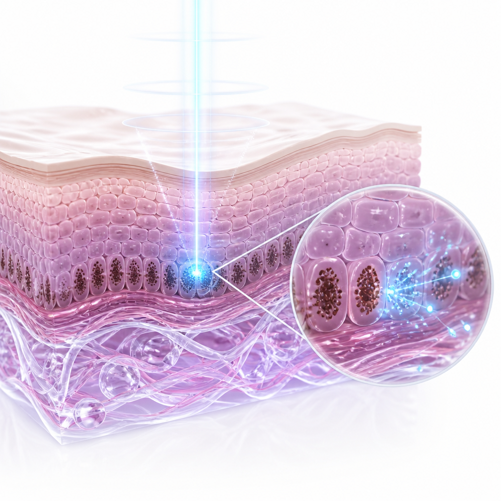
How Do Clinical Laser Systems Compare to Topical Treatments for Areola Whitening?
When addressing congenital areolar hyperpigmentation, choosing the appropriate modality is critical to achieving satisfactory outcomes while preserving tissue integrity. Historically, topical bleaching agents were the primary recourse, but their efficacy on congenital, deep-seated pigment is highly limited. Modern gynecology and aesthetic medicine utilize specialized energy-based devices (EBDs) to achieve precise target-specific photothermolysis. According to multiple observational studies and meta-analyses published in the Journal of Dermatologic Surgery (2021), selective laser systems outperform topical regimens in both the speed of clearance and the longevity of results for sensitive-area hyperpigmentation.
The primary laser systems utilized for areola and intimate whitening are Q-switched Nd:YAG lasers (operating at 1064 nm) and advanced Picosecond lasers. These devices deliver ultra-short pulses of energy that target melanin granules within the skin. The Nd:YAG laser relies on a photothermal and photoacoustic effect to shatter melanin into microscopic particles that are subsequently cleared by the body\’s lymphatic system. Picosecond lasers, on the other hand, utilize primarily photoacoustic pressure to fragment the pigment without generating excessive heat, which significantly reduces the risk of thermal damage to the surrounding delicate epidermis. The table below outlines the primary differences, clinical efficacy, and limitations of these standard approaches.
| Treatment Modality | Primary Mechanism | Efficacy on Innate Pigment | Clinical Advantages | Primary Limitations |
|---|---|---|---|---|
| Picosecond Laser (755nm/1064nm) | Photoacoustic fragmentation of melanin particles | High (Rapidly shatters deep, dense congenital pigment) | Minimal thermal damage, lower risk of PIH, faster recovery | Requires multiple sessions for optimal clearance |
| Q-Switched Nd:YAG Laser (1064nm) | Photothermal and acoustic disruption of melanosomes | Moderate to High (Gradual pigment reduction over time) | Well-established safety profile, highly controllable depth | Moderate heat accumulation; mild temporary erythema |
| Medical Topical Agents (Hydroquinone/Retinoids) | Inhibition of tyrosinase enzyme and cellular turnover acceleration | Low to Moderate (Fails to penetrate deep dermal pigment effectively) | Non-invasive, easy self-application at home | Risk of contact dermatitis, prolonged treatment duration (months) |
According to official clinical consensus guidelines from the Korean Academy of Aesthetic Medicine and international dermatological societies, successful treatment of sensitive-area hyperpigmentation requires combining customized energy parameters with strict post-procedure barrier restoration to minimize the risk of reactive melanocyte activation.
While laser toning offers highly predictable outcomes, clinical caution is always paramount. In patients with Fitzpatrick skin types IV through VI (darker skin phototypes), the epidermal melanin content is naturally higher, which increases the affinity of the laser energy to the epidermis rather than the target pigment. This necessitates a highly conservative approach with lower fluences and extended intervals between treatments to avoid triggering post-inflammatory hyperpigmentation (PIH). However, outcomes may differ in exceptional cases such as patients with underlying endocrine disorders (such as Addison\’s disease or severe polycystic ovary syndrome) that biochemically drive systemic melanocortin receptor stimulation, rendering localized laser therapies less effective unless the primary systemic condition is medically managed first.
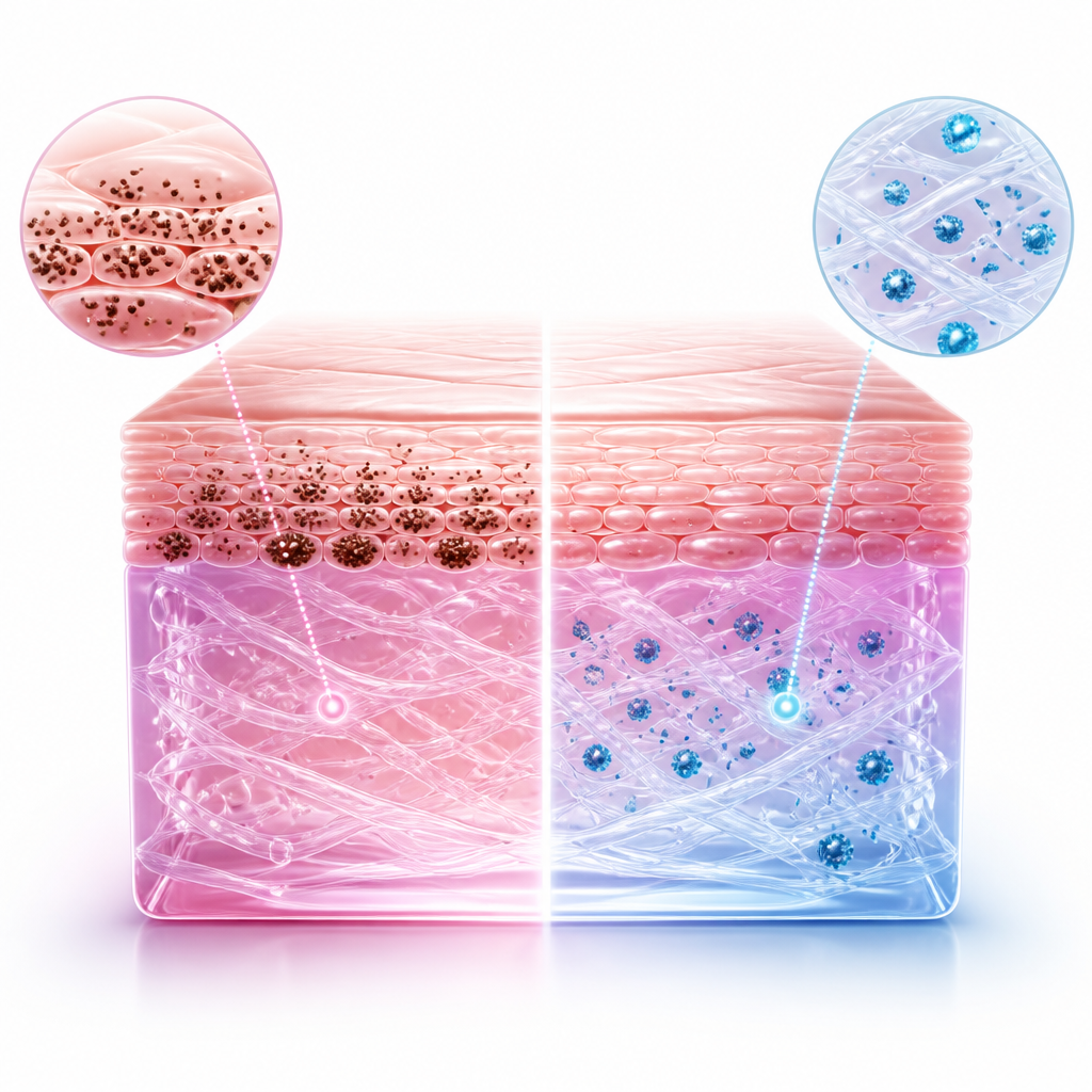
What Criteria Must You Consider Before Choosing a Gynecological Whitening Procedure?
Before proceeding with areola or intimate whitening, a comprehensive pre-treatment evaluation is essential. Because these areas are intimately linked to female reproductive anatomy and hormonal fluctuations, consulting with a specialist in a gynecology-focused setting offers distinct advantages. Gynecological specialists can evaluate whether the hyperpigmentation is purely congenital or if it has been exacerbated by undiagnosed hormonal imbalances, pregnancy-related melasma, or medical conditions affecting the endocrine pathway.
When evaluating a patient for clinical whitening, specialists look at several physiological and lifestyle markers to design a safe, tailored treatment protocol. Patients must be fully informed of the physiological realities of the procedure. To assist in your clinical decision-making process, consider the following key assessment checklist:
- Fitzpatrick Skin Phototype Assessment: Determining your baseline skin phototype is critical to selecting the correct laser wavelength and energy fluence to prevent post-inflammatory hyperpigmentation (PIH).
- Hormonal Stability Verification: Ensuring that your endocrine markers are stable, as active hormonal shifts (e.g., pregnancy, oral contraceptive initiation, or active hormone therapy) can trigger immediate re-pigmentation.
- Tissue Health and Integrity: Evaluating the local tissue for active eczema, contact dermatitis, or localized infections that must be fully resolved before applying any laser energy.
- Expectation and Goal Alignment: Understanding that congenital pigment reduction is a gradual process requiring a series of scheduled treatments, not an overnight modification.
- Commitment to Post-Care Protocols: Acknowledging the absolute necessity of strict UV protection, avoidance of friction, and application of specialized post-procedure barrier creams.
To help navigate the clinical pathway for areola and intimate whitening, the following three-step decision-making flow represents the standard medical approach to patient care:
Step 1 (If congenital hyperpigmentation is confirmed with stable hormones): Initiate a low-fluence, multi-session laser toning protocol using a Picosecond or Q-switched Nd:YAG laser system to safely fragment deep-seated melanin granules.
Step 2 (If the patient presents with dry, sensitive skin or Fitzpatrick Type V-VI): Integrate a pre-treatment skin-conditioning regimen containing mild tyrosinase inhibitors for 2 to 4 weeks prior to the first laser session to stabilize melanocyte reactivity.
Step 3 (If active skin irritation or hormonal fluctuation is present): Postpone energy-based interventions and focus on gentle, non-irritating topical barrier repair until the skin barrier has completely healed and systemic hormones have stabilized.
Frequently Asked Questions FAQ
QIs laser areola whitening safe for future breastfeeding or pregnancy?
Yes, laser areola whitening is clinically safe and does not affect future breastfeeding or pregnancy. The laser energy targets melanin pigments located strictly in the epidermal and upper dermal layers of the skin. It does not penetrate deeply enough to reach or damage the underlying mammary glands, lactiferous ducts, or cooperative sensory nerve pathways. However, it is highly recommended to undergo this treatment after you have completed your family-planning and lactation periods. The significant hormonal surges during pregnancy and breastfeeding naturally stimulate melanocytes, which could easily cause the hyperpigmentation to recur, thereby negating the results of your previous treatments.
QHow many sessions are typically required to see visible results for congenital dark areolas?
Because congenital hyperpigmentation involves genetically active melanocytes that have deposited pigment deep within the skin over many years, a single session is rarely sufficient. On average, a standard clinical protocol requires between 5 to 10 sessions, spaced approximately 3 to 4 weeks apart. This gradual approach is designed to safely break down the melanin layers without causing thermal injury to the delicate skin of the areolar-nipple complex. Most patients begin to notice a gradual brightening and a more even, refined skin tone after the third or fourth session, with compounding improvements continuing throughout the treatment cycle.
QDoes the procedure cause severe pain, and what is the recovery period?
The procedure is highly tolerable and is classified as a minimally invasive, walk-in, walk-out treatment. Before the laser application, a medical-grade topical numbing cream is applied to the treatment area for approximately 20 to 30 minutes, which reduces discomfort to a very mild, warm snapping sensation. Immediately following the session, the skin may exhibit mild redness, slight swelling, or a temporary darkening of the pigment, which typically subsides within 3 to 7 days. There is no disruption to daily life, though patients are advised to wear loose-fitting, soft cotton clothing and avoid vigorous exercise, hot baths, and saunas for the first few days to protect the healing epidermal barrier.
This content is general medical information, and individual treatment decisions should be made through imaging tests and in-person medical evaluation.
Author: Medical content editor based on medical information research
Reviewed by: Specialist consultation from the relevant department
Last reviewed: 2026-07-23
Reference guideline: Korean Academy of Aesthetic Medicine Clinical Protocols 2023
Medical neutrality and closing note
The core of medical decision-making is not to follow a specific device or a trending procedure, but to choose an option that fits each patient’s individual anatomy, condition, risk level, and treatment goals. Every procedure has both advantages and limitations, so decisions should be made after sufficient discussion with an experienced specialist.
[Medical information and copyright notice]
This content is a professional medical column prepared based on medical consultation from 다움산부인과.
The infographics used in this article are created to support understanding and may differ from actual clinical results.
The information provided is a general medical guideline, and accurate diagnosis and treatment require an in-person evaluation by a qualified specialist.